vertebral artery stenosis symptoms
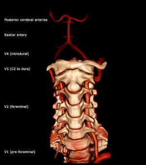
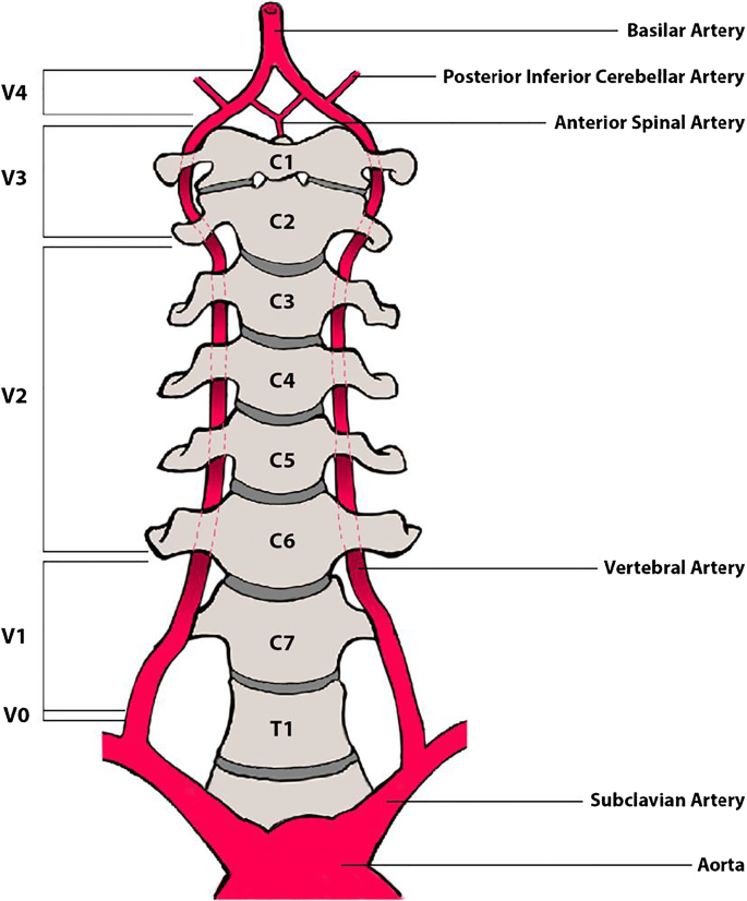
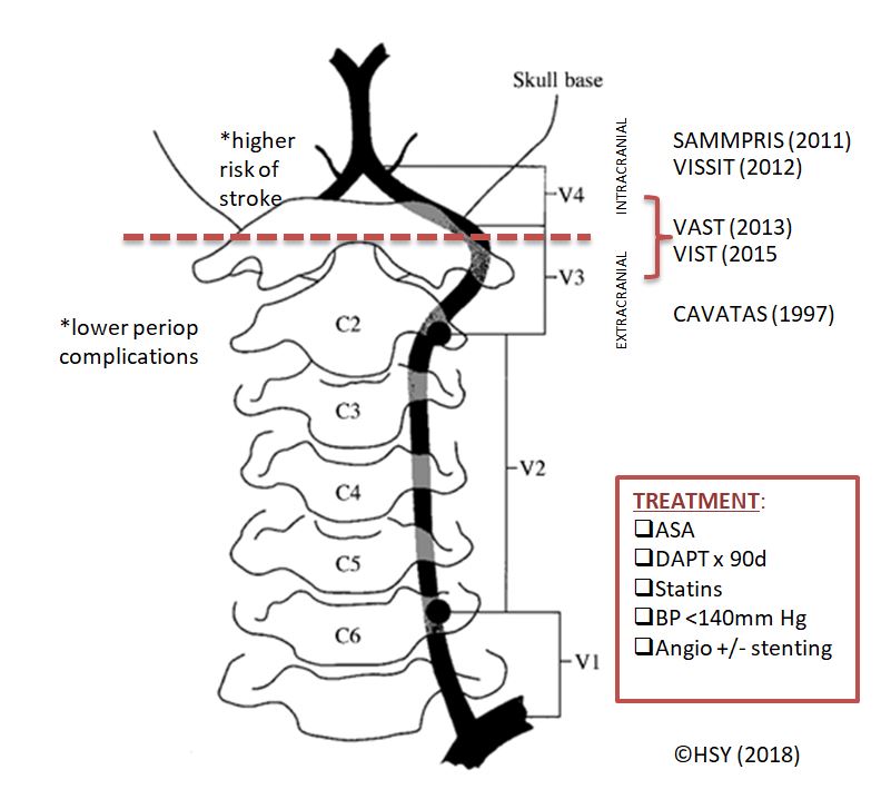
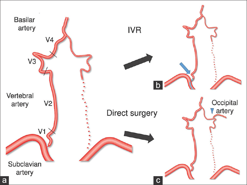
Warning: The NCBI website requires JavaScript to operate. Bilateral Hospital of the vertebral artery present with vertigoDilcan Kotan1Department of Neurology, Faculty of Medicine of the University of Sakarya, Sakarya, TurkeySaadet Sayan2Department of Neurology, SB Sakarya University Education and Research Hospital, Sakarya, TurkeyBilgehan Atilgan Acar2Department of Neurology, SB Sakarya University Education and Research Hospital Ischaemia of vertebral arteries can emerge for different vascular pathological reasons, in different locations and with different clinical findings. Despite their low risk of morbidity and mortality, early diagnosis and treatment is of importance. Symptoms of vertebrobasilar ischemia can be seen clinically as vertigo, tinnitus, double vision, headache, hypokinesis and hearing disorders, etc. This article presents a 42-year-old patient case, which was applied to the emergency service with vertigo and was then diagnosed with bilateral spinal artery stenosis by CRI. BackgroundThe cerebral arteries appear immediately before the thyrocervical trunk on both sides of the neck as two branches of both subclavian arteries; they combine to form the basilar artery at the base of the skull by extending through a neuronal cavity between the cervical vertebrae 1 and 7. The occlusions of the post-brobasilar or vertebrobasilar system are responsible for a quarter of the ischemic blows. The atherosclerosis of the vertebral arteries is the main cause of subsequent circulation afarts. Verbrobasilar ischemia may occur in combination with symptoms such as vertigo, tinnitus, double vision, headache, drowsiness and hearing disorders. Verbral arterial stenosis is one of the recoverable causes of strokes in the back system. Although the risk of morbidity and mortality is low, early diagnosis and treatment is important. In recent years, the number of patients diagnosed and treated has been increasing according to advances in medical imaging methods. This study presents the case of a man with young vertigo and stroke, diagnosed with bilateral stenosis of the vertebral artery. Case presentation A 42-year-old hypertensive man consulted the emergency service with headache and vertigo complaints. When admitted, his neurological examination was normal, except for the left nistagmus with a horizontal rapid rotating phase and minimal central facial palliative on the right side. He was admitted to the hospital as a patient at the time of seeing concordant hypodemic area with infarction in different bilateral cerebellons on the right and in the talamo on the left side (A,B) in the CT scan. Heparin with low molecular weight and 300 mg/day of acetylsalicylic acid twice a day was given to the patient during the hospitalization period. Ramipril 10 mg/day was included in treatment with high blood pressure reasons. On the second day of hospitalization, the right central facial paralysis, mild disarray, left dysmetria, left dysdiacokinesia and left truncale ataxia were present in the patient's examination with advanced neurological findings. Pervasive and diffusive infart areas were observed in the bilateral cranial cerebellege of the MRI, the left occipital, the right mesencephalon and the left thalamic zone (A-C). A restriction on dissemination in these areas was observed in the dissemination of MRI (A,B). It was impossible to see the right vertebral flow in the Doppler ultrasound (USG) the examination of the carotis vertebral arteries and 70% of stenosis was detected in the left vertebral artery. As a result, in the expected vertebral MR angiography, distal occlusion was observed in the intermediate and distal sections of the right vertebral artery and distal distal of the left vertebral artery (A,B). Balloon angioplasty was applied to the patient diagnosed with bilateral stenosis of vertebral arteries.(A and B) Hypodermic area concordant with infarction in a bilateral cerebelous zone different on the left side (A) and in talamo on the left side in the CT scan.(A-C) Debate The roots of the vertebral artery of the proximal subclavian. The vertebral artery reaches the cervical vertebrae after passing through the transversal foramenes of the fifth and sixth vertebral after leaving the subclavian artery. The segment outside the form 'V1', the cervical segment within cross-sectional 'V2', segment to the point where subarachnoid space enters after passing through the 'V3' hard-to-matter 'V4' segment after entering the subarachnoid space, and forms the basilar artery by combining 'V4' in the front of the bulb. Ischaemia of vertebral arteries may occur due to different vascular pathological causes, in different places and in different clinical findings. Verbral arterial stenosis is accompanied by a wide range of findings from the back system of a simple vertigo to the dysfunction of the occipital cortex (vertigo, loss of balance, ataxia, tinnitus and gout attacks). When our patient was admitted to the hospital, there was only one symptom that was vertigo, and 2 days later, ataxia and other cerebellon findings were involved. In the images of the emergency tomography, it was monitored closely and an anticoagulant treatment was initiated due to the presence of suspected infarctions. Since the vertebral artery system has a structure that can be considered complex, the evaluation of stenosis with ultrasonography (USG) with Doppler color can be difficult. It is known that a good image quality, a high level of technical knowledge and experience are all necessary in the evaluation of stenosis with ultrasonography with Doppler color. RM Vertebral's angiography has been introduced as a common practice in recent years and as a complementary method for obtaining detailed information on vascular pathology. In our case, it was impossible to see the right vertebral flow with the USG with Doppler color and the flow rate of the left vertebral decreased significantly. The angiography of the vertebral artery revealed that intermediate and distal sections of the right and left vertebral distal were occluded. In our case, the USG with Doppler color and angiography of the MR vertebral artery suggested the same vascular pathology. Antitrombotic and anticoagulant drugs were used to reduce the risk of stroke in vertebral artery stenosis. In our patient, anticoagulant treatment began from the early stages. Interventional treatment was considered, as clinical findings were progressive despite treatment. Surgical treatment is avoided in spinal artery stenosis due to frequent occurrences of complications such as Horner syndrome (15–28%), lymphocytes (4 percent), recurrent laryngeal nerve lesion (2%). In cases where symptoms are persistent after proper medical treatment, there is an indication for treatment with angioplasty and ball stent. However, there is no other study to compare the success rates of two methods. Our case was admitted to the neuroradiology department, bearing in mind that endovascular intervention would be appropriate and a balloon angioplasty was applied. Learning point As a result, the pathologies of vertebral artery should be taken into account in individuals with central system-related vertigo, regardless of the presence of vascular risk factors. Footnotes Comparison Interests: None. Consent of the patient: Obtained. Inter-couple provenance and examination: Not in charge; peer review has been conducted externally. ReferencesFormats: Share , 8600 Rockville Pike, Bethesda MD, 20894 USA
Vertebral Artery Test - Physiopedia

Stenting versus medical treatment in patients with symptomatic vertebral artery stenosis: a randomised open-label phase 2 trial - The Lancet Neurology

Treating Vertebrobasilar insufficiency – Bow hunter's syndrome – Caring Medical Florida

Endovascular Treatment of Vertebral Artery Stenosis - ScienceDirect
Vertebral Artery Stenosis | SpringerLink
Vertebral Artery Stenosis Studies – Peripheral Brain

Subclavian Steal Syndrome Presentation and Treatment | medcaretips.com

Vertebral Artery Injury - Spine - Orthobullets

Endovascular Treatment of Vertebral Artery Stenosis - ScienceDirect

Patient circulation with Saltzmann type 2 PTA and internal carotid... | Download Scientific Diagram
Occipital artery to extracranial vertebral artery anastomosis for bilateral vertebral artery stenosis at the origin: A case report - Surgical Neurology International

Rotational Vertebral Artery Occlusion | Stroke
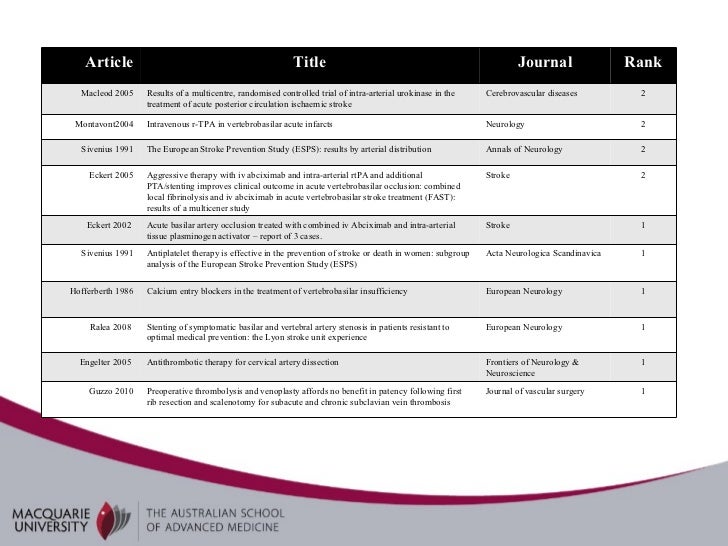
Vertebral artery stenosis
Vertebrobasilar Artery Occlusion - The Western Journal of Emergency Medicine
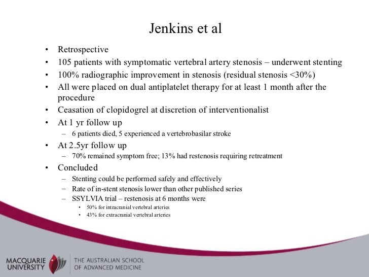
Vertebral artery stenosis

Clinical manifestations of vertebral artery dissection. | Download Table
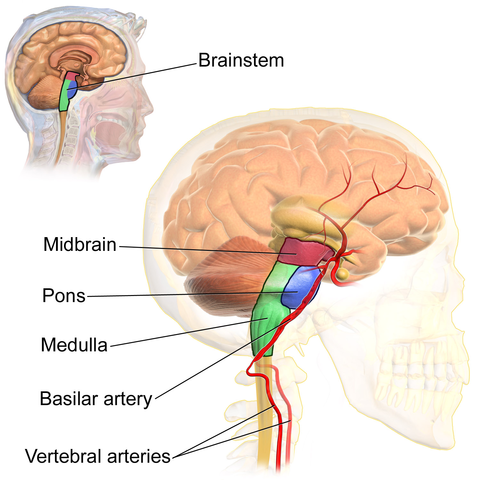
Anatomy and Diseases of the Vertebral Artery | Lecturio
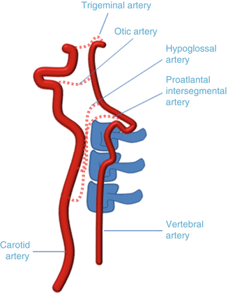
Imaging of the Pathology of the Vertebral Arteries | Radiology Key

Vertebral PTA: Indications and Technique Patrick L. Whitlow, MD Director, Interventional Cardiology The Cleveland Clinic Foundation Patrick L. Whitlow, - ppt download

Vertebral artery dissection - Wikipedia
The crucial, controversial carotid artery Part I: The artery in health and disease - Harvard Health
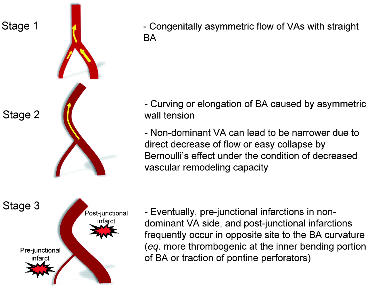
Vertebral artery dominance contributes to basilar artery curvature and peri-vertebrobasilar junctional infarcts | Journal of Neurology, Neurosurgery & Psychiatry
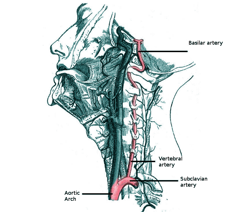
Vertebrobasilar Insufficiency Article
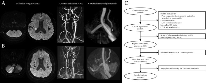
Long-term outcome of vertebral artery origin stenosis in patients with acute ischemic stroke | BMC Neurology | Full Text

Ultrasound Assessment of the Vertebral Arteries | Radiology Key

Stenting for symptomatic vertebral artery stenosis: a preplanned pooled individual patient data analysis - The Lancet Neurology
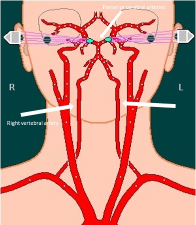
Transient Vertebrobasilar Insufficiency - SeattleNeurosciences.com
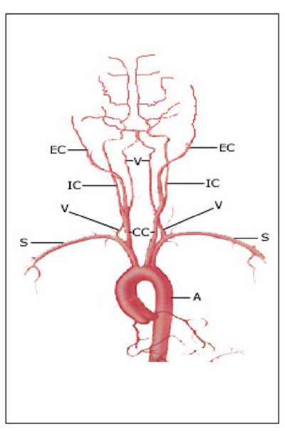
Visual Problems Result From Severe Stenosis

Stroke mechanisms and outcomes of isolated symptomatic basilar artery stenosis | Stroke and Vascular Neurology

Subclavian steal syndrome - Wikipedia
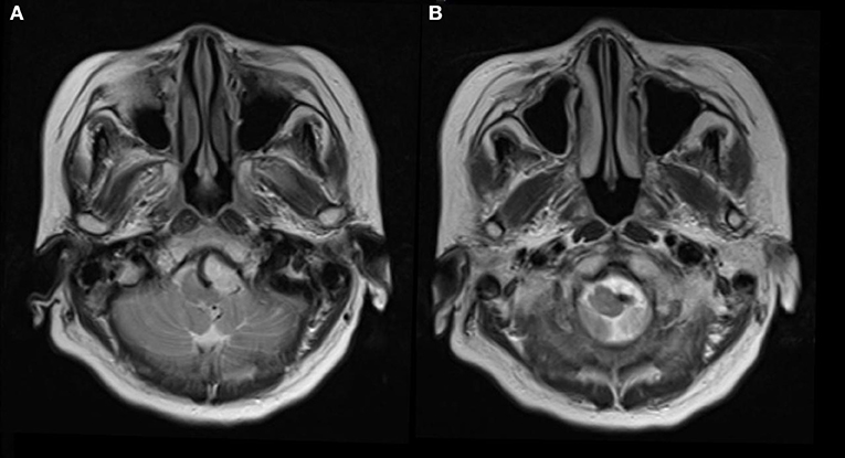
Frontiers | Vertebral Artery Compression Syndrome | Neurology

Dizziness due to Subclavian Steal
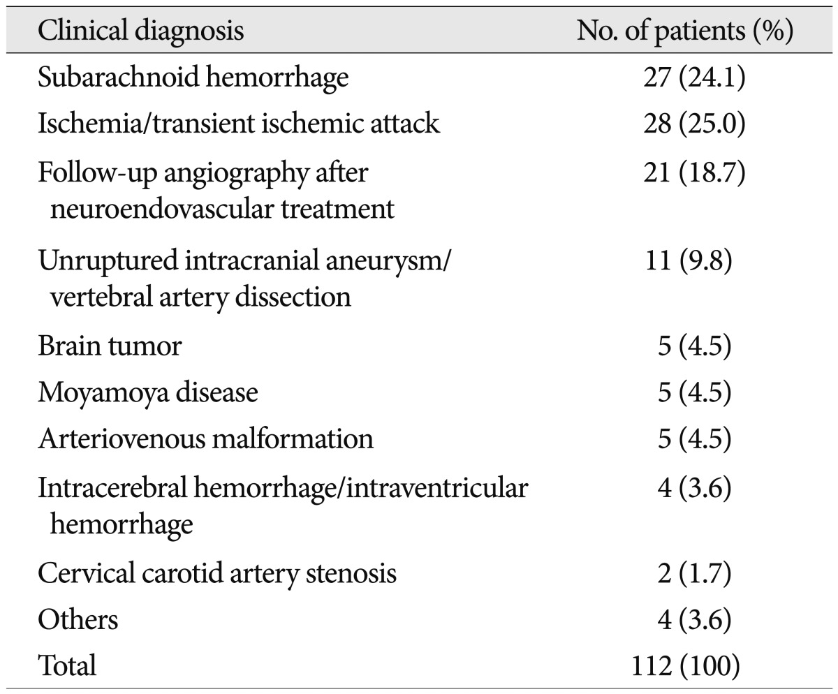
Journal of Korean Neurosurgical Society
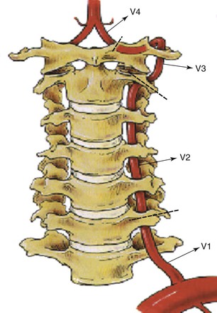
Vertebral Artery Disease | Thoracic Key
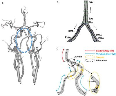
Frontiers | Vertebral Artery Stenoses Contribute to the Development of Diffuse Plaques in the Basilar Artery | Bioengineering and Biotechnology

Subclavian Artery Disease: Diagnosis and Therapy - The American Journal of Medicine
Stenting for symptomatic vertebral artery stenosis

A) Hypoplastic right vertebral artery (bottom arrow); basilar artery... | Download Scientific Diagram

Subclavian Steal Syndrome | Circulation

Clinical Investigation and Characterization of Vertebrobasilar Dolichoectasia and Vertebral Artery Dominance - Xiangfang Meng - Discovery Medicine
Posting Komentar untuk "vertebral artery stenosis symptoms"